(II) Routine Resolution AFM
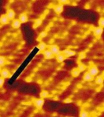
The image to the right shows the surface of a crystal of benzoin (C6H5CHOHCOC6H5) as it dissolves under a liquid of 95% water : 5% n-propanol (as if we're looking at the bottom of a shallow pond.
The 256x256 pixel image spans about 5 microns (1/10th of a hair's breadth). Each benzoin molecule is about 5Ċ (0.5 nm) wide. How many molecules wide is the picture? How many molecules wide is a pixel ?
The surface is colored as if illuminated by a light from the left (like a satellite picture taken with afternoon sun). Two light, straight lines crossing the whole image are steps separating terraces that differ in height by two molecules. Actually two single-moledule steps join near the top left to make the top step double along most of its length.
The AFM tip has been used to scratch a cross about 10 molecular layers deep near the center of the image.
[This is one frame from a movie showing how the crystal dissolves. The movie was made at Yale by Kraig Steffen Click to see the movie (364K).]
(III) Near-Atomic Resolution STM
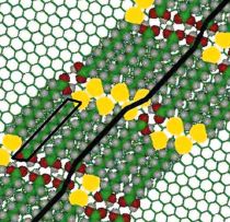
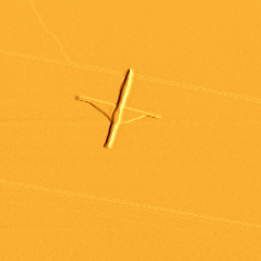
Here is an STM image showing some 25 molecules of 12-bromoundecanoic acid [Br(CH2)12COOH] lying down side-by-side in rows to make a layer one molecule thick on the flat surface of graphite. Below the STM image is a model diagram explaining what it shows.
The black bar marks the location of a single long molecule stretching some 1.5 nm (15 Ċ) from the dark brown COOH group on the lower left to the bright yellow Br atom on the upper right.. A corresponding molecule is outlined in a box at the lower left of the artist's model of the packing shown below. (Note that successive molecules along the row traced by the long black line are anti-parallel, meeting COOH to COOH then Br to Br.)
The molecules are sufficiently thin that electrons can "tunnel" down through them from the sharp(!) metal tip to the electrically conductive graphite (which is sketched as a pattern of hexagons at corners of the model where layer of acid molecules has been removed. The extreme sensitivity of "tunnelling" current to distance, when the tip is almost touching the molecule being imaged, allows clear, single-molecule resolution without damaging the delicate monolayer. The image almost has atomic resolution, since, knowing what to look for, one can trace the zig-zag chain of 11 light brown carbon atoms (green in the model) between the COOH group and the bromine atom, and perhaps see the H atoms as small yellow balls (gray in the model).
[For this image we thank Dalia Yablon and the Columbia University research lab of Professor George Flynn (Yale '60). More images are available on the group's web page.]